From Injury to Recovery:
The Role of Visualization in Trauma Reconstruction
May 14th, 2026
By Kloie Bautz
Trauma reconstruction is inherently complex.
Unlike planned procedures, trauma cases rarely follow a predictable path. Surgeons are often working through disrupted anatomy, variable tissue viability, and time-sensitive decisions—all at once.
In those moments, having clear visibility into the surgical field becomes essential.
Because within that complexity, identifying and navigating critical structures can shape how reconstruction unfolds—and nerves, in particular, present a unique challenge.
Why Nerve Preservation Matters
Nerves are fundamental to function.
They enable movement, sensation, and coordination—systems that patients rely on long after the surgical procedure is complete. In trauma settings, damage to these structures is often described as
peripheral nerve injury, which can significantly affect both motor and sensory function.
When nerves are damaged or unintentionally injured during surgery, the effects can extend well beyond the operating room. Loss of sensation, impaired motor control, and chronic pain are all recognized consequences of peripheral nerve injury. Even when reconstruction is technically successful, functional recovery may remain limited.
This is especially relevant in trauma cases such as:
- Mangled extremity injuries
- Complex soft tissue reconstruction
- Facial trauma
In these situations, preserving nerve integrity is closely tied to long-term patient outcomes.
The Challenge of Visibility in Trauma
One of the core difficulties in trauma reconstruction is visibility.
Anatomy is often altered by the injury itself. Bleeding, swelling, and tissue disruption can obscure important structures, making identification more difficult.
Unlike vessels or bone, nerves can be particularly hard to distinguish within the surgical field.
As a result, surgeons frequently rely on:
- Anatomical knowledge
- Experience and intuition
- Indirect cues within the tissue
While these approaches are effective, they are not always sufficient—especially in high-variability cases.
This creates a
persistent challenge: how can surgeons gain better visibility in the moment without adding complexity to the procedure?
The Role of Visualization in Surgical Decision-Making
Small choices—where to dissect, what to preserve, how to approach reconstruction—can have lasting consequences. The more clearly a surgeon can interpret the surgical field, the more confidently those decisions can be made.
Visualization plays a central role in that process.
Improved visibility does not replace surgical skill. Instead, it supports it—providing additional information at critical moments. In trauma reconstruction, this can influence:
- Identification of viable tissue
- Recognition of nerve pathways
- Decisions around preservation or repair
Ultimately, better visualization has the potential to support both precision and consistency across cases.
Looking Forward: A Shift Toward Function-Focused Innovation
Surgical success isn’t just defined by the procedure itself.
It’s reflected in how a patient recovers in the weeks and months that follow.
In trauma reconstruction, outcomes are often defined by function—movement, sensation, and quality of life.
Decisions made in the operating room play a central role in shaping those outcomes.
At YaYa Scientific, this perspective has shaped the development of
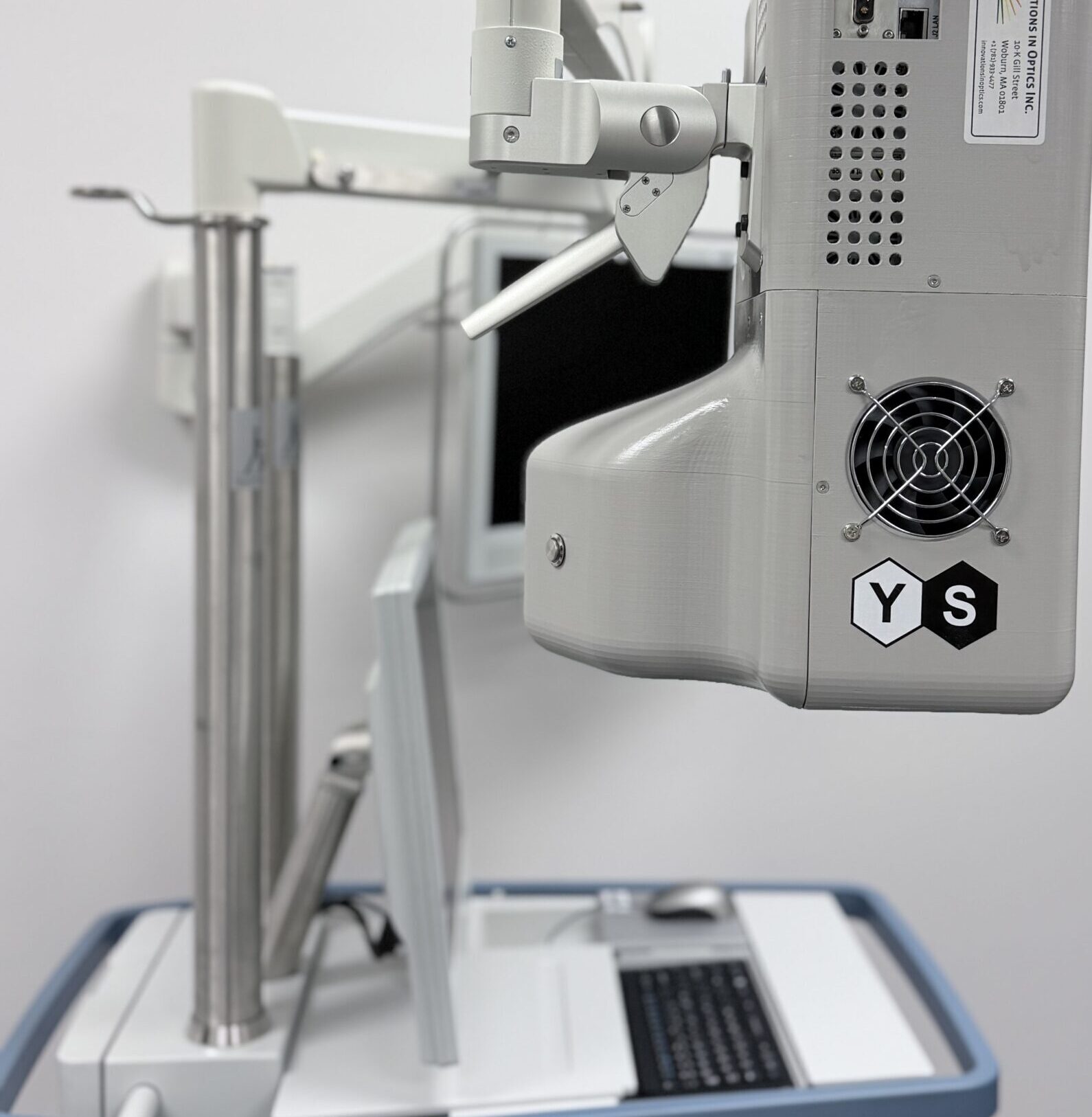
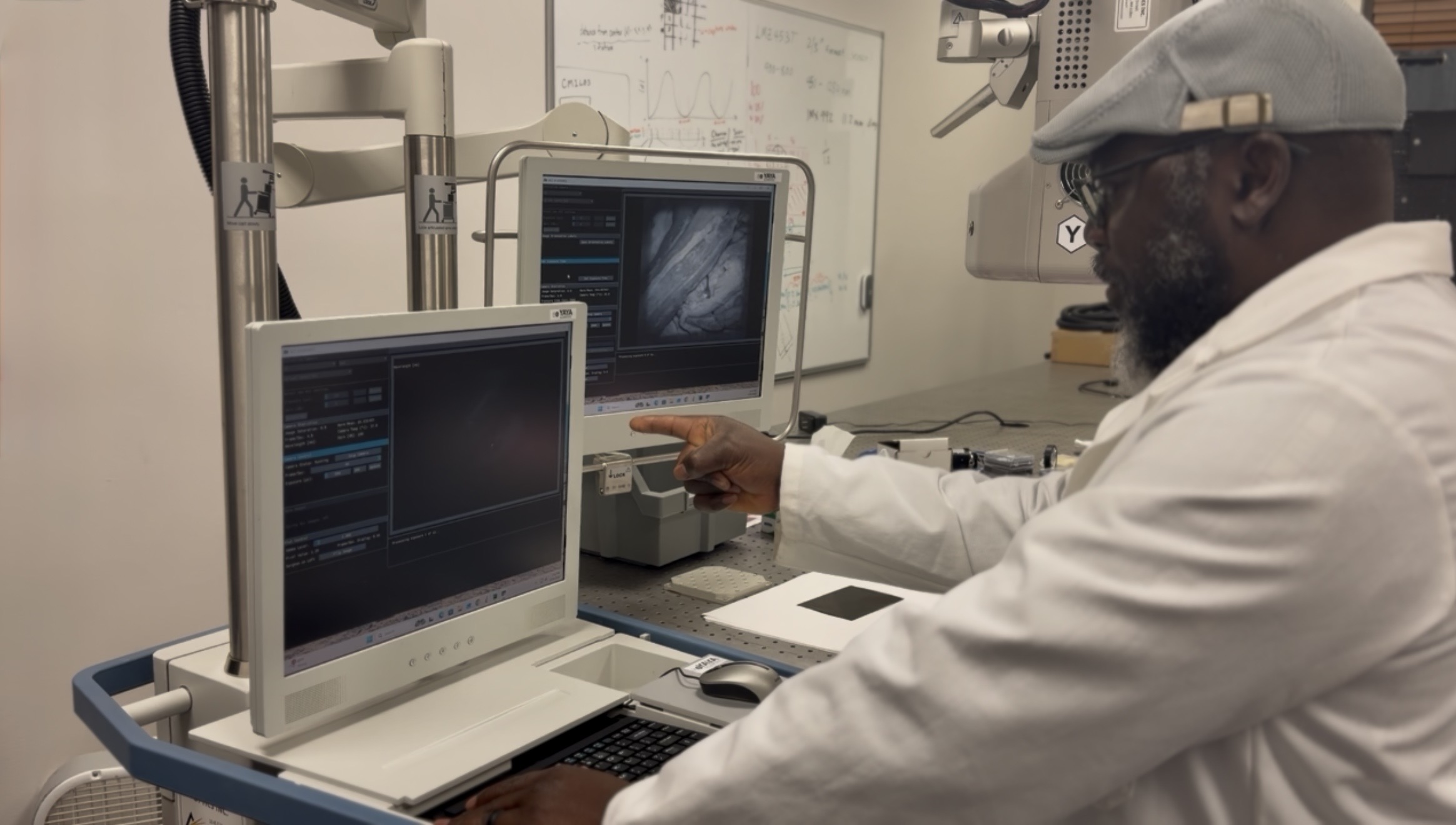
Alina™, a system designed to provide real-time visualization of nerve structures during surgery. It helps surgeons to better see and identify nerves within the surgical field, supporting more informed decisions without disrupting existing workflows.
Conclusion
Trauma reconstruction will likely always involve a high degree of complexity.
But even within that complexity, small improvements in how surgeons visualize and interpret the surgical field can make a meaningful difference.
In cases where nerve preservation is critical to function, those differences can shape recovery long after the procedure is complete.
About the Author
Kloie Bautz, BS BME
Project Engineer
·
YaYa Scientific