June 15th, 2026
Across multiple surgical specialties, the literature continues highlighting a common challenge: identifying and preserving delicate nerve structures in anatomically complex surgical environments.
Although the procedures themselves can differ significantly, many of the underlying challenges remain remarkably consistent.
Scar tissue, bleeding, swelling, fracture displacement, prior surgery, anatomical variability, and limited exposure can all complicate intraoperative orientation and make nerve identification substantially more difficult. In many open procedures, surgeons are operating within narrow visual margins around structures that may only be partially visible during exposure and reconstruction.
These challenges matter because the consequences of nerve injury can be significant for long-term patient function and recovery.
Depending on the procedure and nerve involved, injury may contribute to chronic pain, sensory deficits, weakness, neuropathy, foot drop, voice changes, or long-term functional impairment.
As we continued reviewing these surgical environments, a broader pattern began to emerge.
What became increasingly clear was how often successful nerve preservation depended on visibility and anatomical orientation during surgery.
Orthopedic and trauma procedures are particularly interesting in this context because many involve direct exposure around vulnerable nerve structures during fixation and reconstruction.
In acetabular and hip procedures, for example, the sciatic nerve may become vulnerable depending on surgical approach, retraction, fracture displacement, and surrounding anatomy. In revision procedures especially, scar tissue and distorted anatomy can make orientation significantly more difficult.
Similar patterns appear in humeral fixation procedures, where the radial nerve travels closely along the humerus and may require formal identification and mobilization during surgery.
In distal humerus and elbow procedures, the ulnar nerve is frequently encountered directly during exposure and reconstruction, creating another environment where visualization and nerve awareness remain critically important.
Across many of these procedures, some of the most technically demanding portions of surgery occur during exposure and navigation through anatomy that may only be partially visualized intraoperatively.
Similar themes also appear throughout breast surgery and reconstruction literature.
During axillary lymph node dissection procedures, surgeons may intentionally identify and preserve the intercostobrachial nerve in an effort to reduce postoperative sensory disturbance and neuropathic pain.
At the same time, reconstructive approaches focused on restoring sensation following mastectomy have increased interest in nerve identification and preservation.
These procedures are especially notable because nerve location, branching patterns, and surrounding anatomy can vary considerably between patients, making consistent intraoperative identification more difficult.
Many of the same visualization challenges also appear in open spine surgery and other deep exposure procedures.
Whether operating around nerve roots, thecal structures, or anatomically constrained surgical spaces, visualization and orientation remain central to safe surgical workflow. In these environments, even small inaccuracies around delicate neural structures can contribute to pain, weakness, sensory deficits, or long-term functional complications.
Even as minimally invasive and robotic-assisted approaches continue expanding in some specialties, many procedures involving trauma reconstruction, fixation, decompression, and microsurgical reconstruction still rely heavily on open surgical exposure.
Across all of these environments, the importance of visualization continues appearing repeatedly throughout the literature.
What has become increasingly apparent throughout this research is how many surgical procedures already depend heavily on careful nerve identification and preservation.
Despite involving very different anatomy and surgical approaches, many of these specialties continue facing remarkably similar challenges surrounding visibility, anatomical orientation, and nerve preservation.
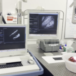
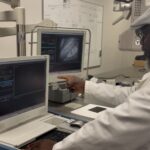
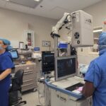
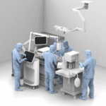
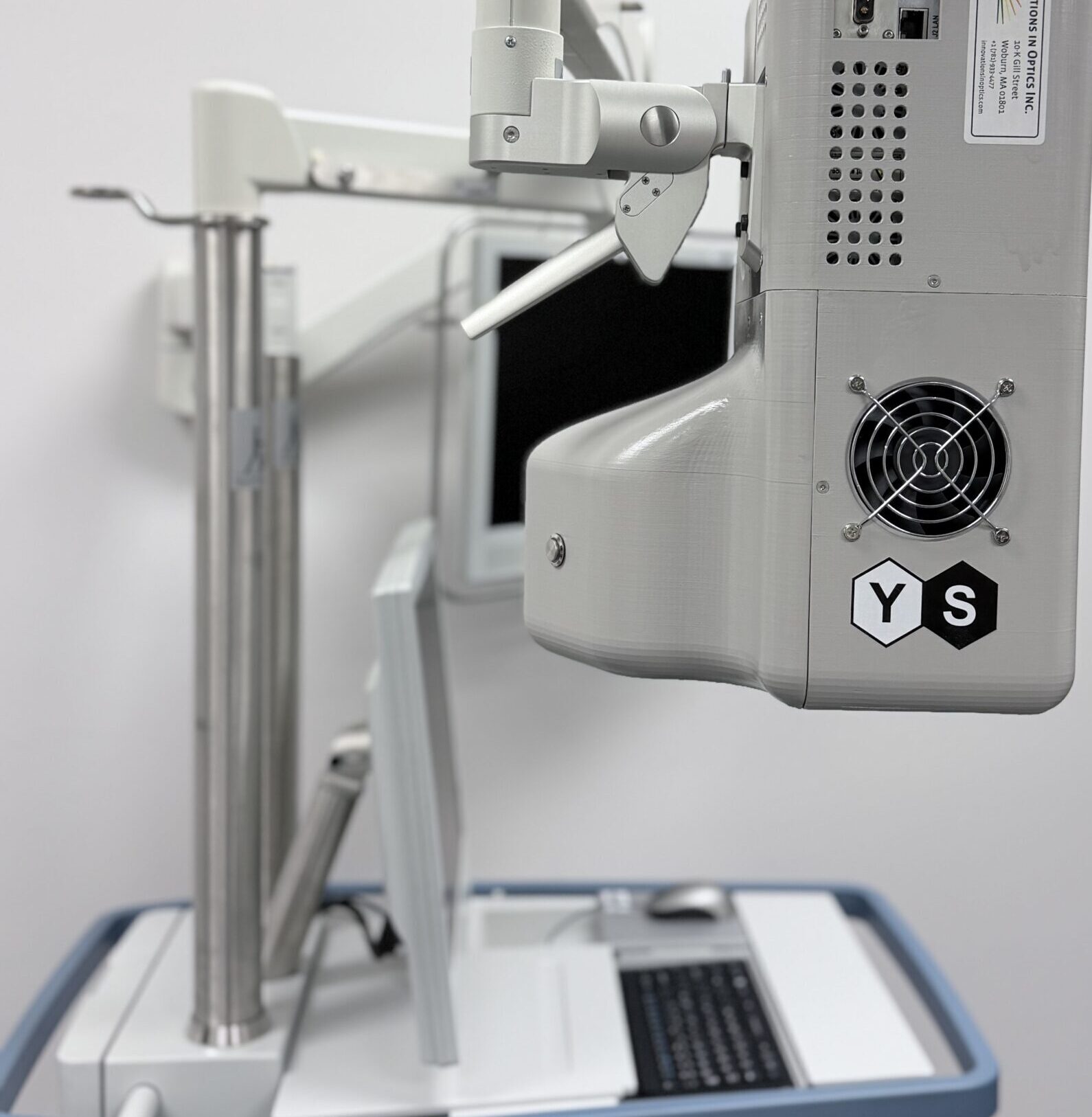
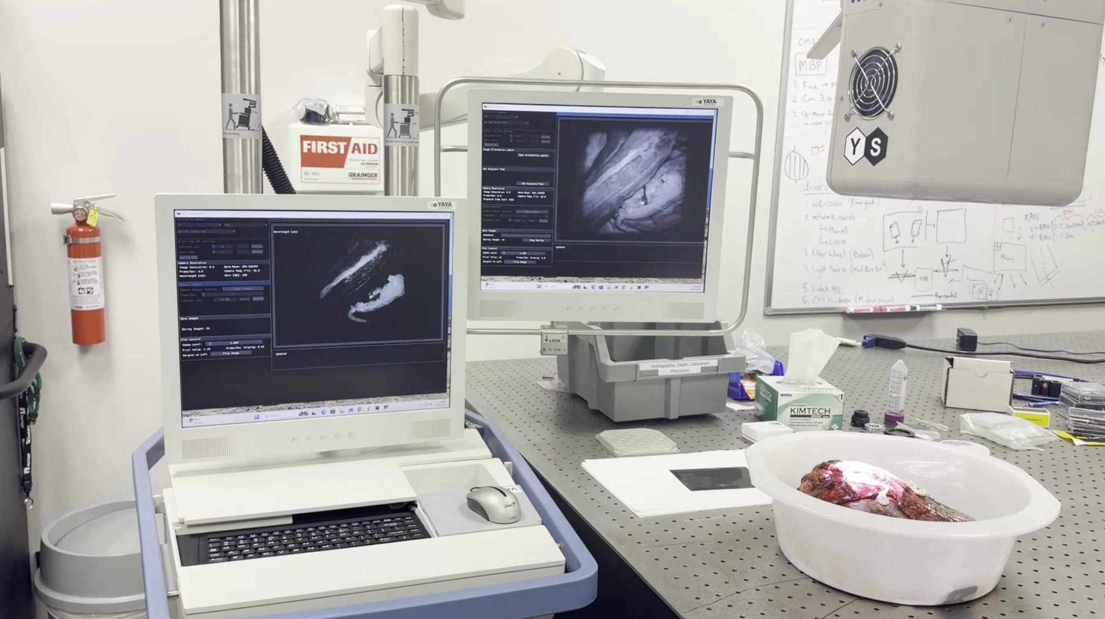
Current development work around the Alina™ platform remains focused on thyroid surgery.
However, studying these broader surgical environments continues to shape how we think about the future role of surgical visualization technologies across open procedures and where improved intraoperative visualization may eventually support nerve identification in inherently complex surgical settings.
The Alina™ platform is currently in development and is not FDA approved. Current development work is focused on thyroid surgery applications.